About the project
Objective
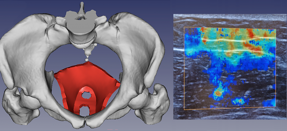
We develop a novel multimodal imaging database, PelvicMIM, by integrating next-generation digital diagnostic technologies to advance the evaluation of childbirth-related pelvic floor muscle injuries. This effort includes the development and validation of cutting-edge imaging modalities—Shear Wave Elastography (SWE), Magnetic Resonance Elastography (MRE), and Diffusion Tensor Imaging (DTI). These techniques will be applied in vivo to quantify the biomechanical and structural properties of pelvic floor muscles. A deep learning-based image processing framework will be designed for multimodal image registration, enabling the overlay of stiffness maps from MRE/SWE and fiber orientations from DTI onto MRI and ultrasound images. Our proposed approach facilitates cross-modality findings, offering deeper insights into muscle function and injury mechanisms.
Background
One in two middle-aged women suffer from pelvic floor dysfunction such as urinary and fecal incontinence or prolapse of the pelvic organs into the vagina, which profoundly impair quality of life. Injuries to the pelvic floor muscles due to birth are highly associated with pelvic floor dysfunction later in life. Nevertheless, injuries to these muscles, which cannot be surgically repaired, have been largely ignored and poorly studied. The Swedish Agency for Health Technology Assessment, SBU, has identified birth-related injuries to the levator ani muscle (LAM), the three largest muscles of the pelvis, as a priority area for research (April 2019). Although recent research also highlights the urgent need for quantitative assessment of LAM injuries, clinical practice still relies on conventional ultrasound, which lacks the ability to quantify biomechanical or structural properties that are important indicators of soft tissue health. These properties are crucial for the assessment of the LAM, as it is a complex structure of three muscles working together in a sheet-like shape with different layers and fiber directions.
Crossdisciplinary collaboration
The team of researchers is composed of members from the KTH School of Engineering Sciences in Chemistry, Biotechnology and Health, Department of Biomedical Engineering and Health Systems and KTH School of Engineering Science, Department of Engineering Mechanics. The project is conducted in close collaboration with clinical partners at Karolinska University Hospital.