About the project
Objective
The purpose of this project is to develop better methods for reconstructing time-resolved medical images with multiple image channels. Traditional methods often process these measurement subsets separately and then combine the results, but this approach doesn’t always lead to the best images. The challenge is to integrate the extra information from the start, during the image reconstruction process itself, in a way that enhances the final result. This is where artificial intelligence (AI) and deep learning come in.
Deep learning models have shown great promise in tackling complex tasks by learning patterns from large amounts of data. However, in medical imaging, data is often scarce, and the computational challenges are significant. A fully data-driven approach is unlikely to succeed. Instead, our project will explore ways to build AI models that incorporate the known relationships between measurement subsets directly into their design. This will allow us to develop efficient and lightweight models that improve image quality while remaining practical to use in real-world clinical settings.
By applying these new methods to spectral CT and PET, we aim to produce medical images with greater diagnostic power, helping doctors detect and treat diseases more effectively. Additionally, our approach will be designed in a flexible, “plug-and-play” manner, so that it can be adapted for other types of imaging in the future. With this research, we hope to take an important step toward more accurate, reliable, and informative medical imaging for patients and healthcare providers alike.
Background
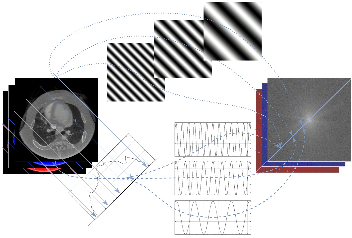
Medical imaging techniques like Computed Tomography (CT) and Positron Emission Tomography (PET) allow doctors to see inside the human body without surgery. However, these images are not captured directly like a photograph. Instead, they are computed from indirect measurements of light particles (photons) passing through the body.
Imagine looking at the shadows cast by an object in different directions and trying to piece together what the object looks like in three dimensions. This is similar to how medical images are reconstructed from projection data. Some examinations build on acquiring multiple image channels, such as in spectral CT where X-ray images are acquired at multiple energy levels, or combining different modalities such as PET and CT. It is also possible to acquire a video sequence of multiple images in rapid succession. Combining these different kinds of information has the potential to improve image quality, making diagnoses more accurate, but how this should be done in an effective way is far from a simple question.
Cross-disciplinary collaboration
Developing novel medical imaging methodology is a highly cross-disciplinary activity which requires involvement of physics, mathematics, computer science, engineering, and medical science. In this project, which is a collaboration between the department of physics (SCI), the department of biomedical engineering and health systems (CBH), and the department of mathematics (SCI) at KTH, we bring together expertise in mathematics and in two different imaging modalities, CT and PET, to develop common methodology that can be applied to multiple medical imaging modalities.